40 label the skin structure and areas indicated in the accompanying diagram of skin
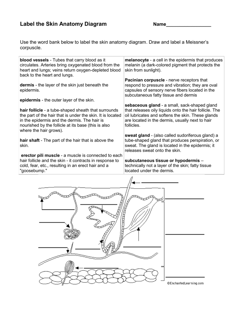
The Integumentary System - Holly H. Nash-Rule, PhD Label the skin structures and areas indicated in the accompanying diagram of thin skin. Then, complete the statements that follow. a. Lamellar granules contain glycolipids that prevent water loss from the skin. b. Fibers in the dermis are produced by fibroblasts . c. 5.1 Layers of the Skin – Anatomy & Physiology 1). The most superficial layer of the skin is the epidermis which is attached to the deeper dermis. Accessory structures, hair, glands, and nails, are found ...
PDF Integumentary System Review Sheet Exercise 7 Answers Basic Structure of the Skin 1. Complete the following statements by writing the appropriate word or phrase on the correspondingly numbered blank: Epidermis The two basic tissues of which the skin...

Label the skin structure and areas indicated in the accompanying diagram of skin
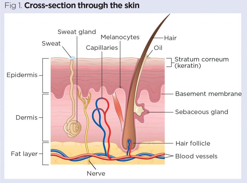
Living Environment - New York Regents June 2009 Exam - Syvum Answer: 13. 13 Carbon dioxide makes up less than 1 percent of Earthâ s atmosphere, and oxygen makes up about 20 percent. These percentages are maintained most directly by (1) respiration and photosynthesis (2) the ozone shield (3) synthesis and digestion (4) energy recycling in ecosystems. Answer: Assignment 11 pg 104.pdf - 4. Label the skin structures and... Label the skin structures and areas indicated in the accompanying diagram of thin skin. Then, complete the statements that follow. Subcutaneous J tissue or _l T~P-r Integumentary System - Building a Medical Terminology Foundation The skin and accessory structures are the largest organ system in the human body. The integumentary system refers to the skin and its accessory structures. In the adult human body, the skin makes up about 16 percent of body weight and covers an area of 1.5 to 2 m 2. In fact, the skin and accessory structures are the largest organ system in the ...
Label the skin structure and areas indicated in the accompanying diagram of skin. Solved Basic Structure of the Skin 1. Complete the following - Chegg Label the skin structures and areas indicated in the accompanying diagram of thin skin. Then areas indicated in the accompanying diagram of thin skin. Then, complete the statements that follow. same Sratum Sratum Sratum. Cell Organelles- Definition, Structure, Functions, Diagram Cell organelles are specialized entities present inside a particular type of cell that performs a specific function. There are various cell organelles, out of which, some are common in most types of cells like cell membranes, nucleus, and cytoplasm. However, some organelles are specific to one particular type of cell-like plastids and cell ... Mitosis & Meiosis | Genetics Quiz - Quizizz answer choices. Cell B contains the same genetic information that cells A and C contain. Cell C has DNA that is only 50% identical to cell B. Cell A has DNA that is only 75% identical to cell B. Cells A, B, and C contain completely different genetic information. Question 4. Untitled - Holly H. Nash-Rule, PhD Name Lab Time/Date The Integumentary System Basic Structure of the Skin 1. Complete the following statements by writing the appropriate word or phrase on the correspondingly numbered blank: The two basic tissues of which the skin is composed are dense irregular 1. connective tissue, which makes up the dermis, and 1 , which forms the epidermis.
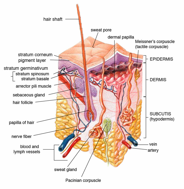
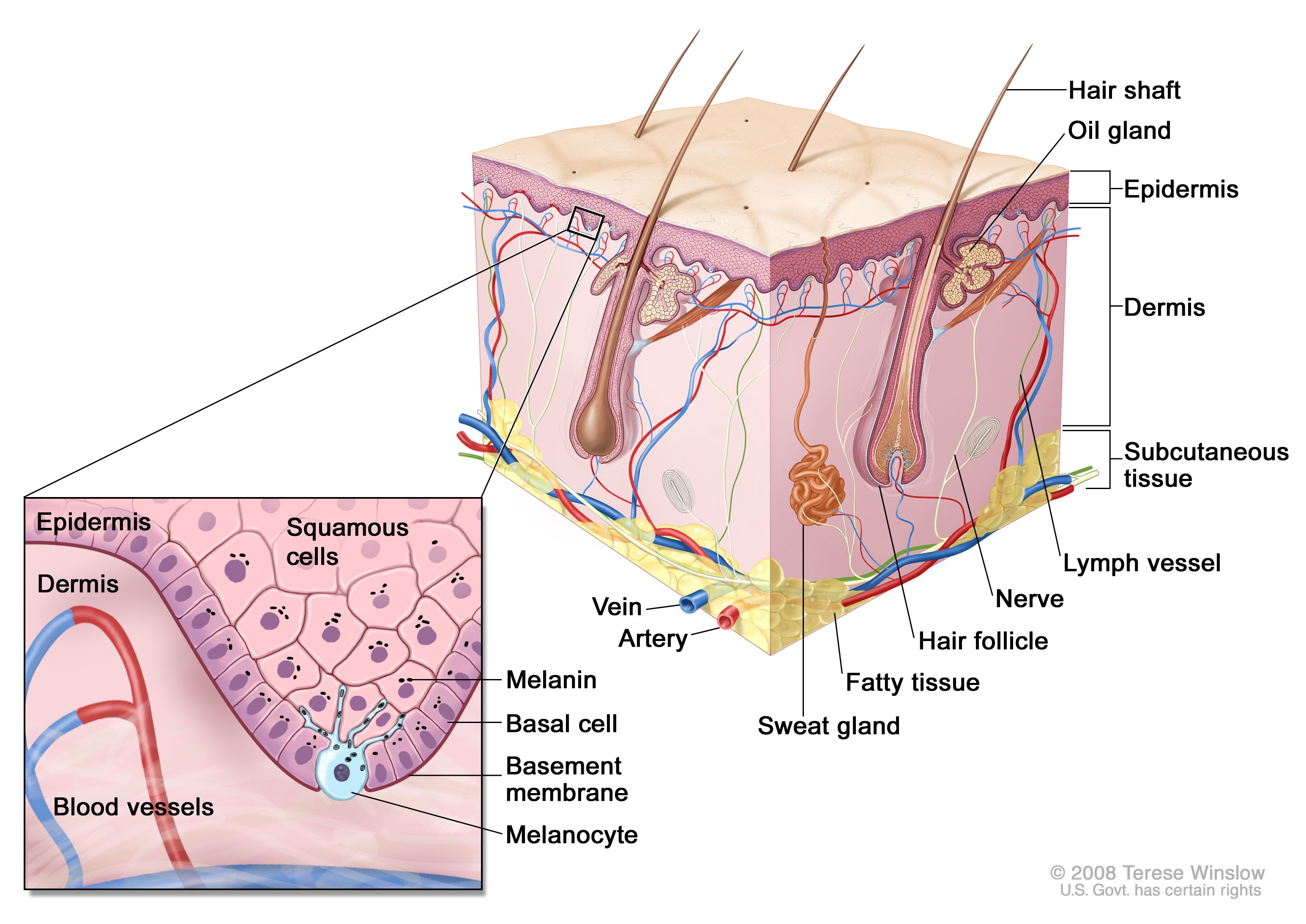
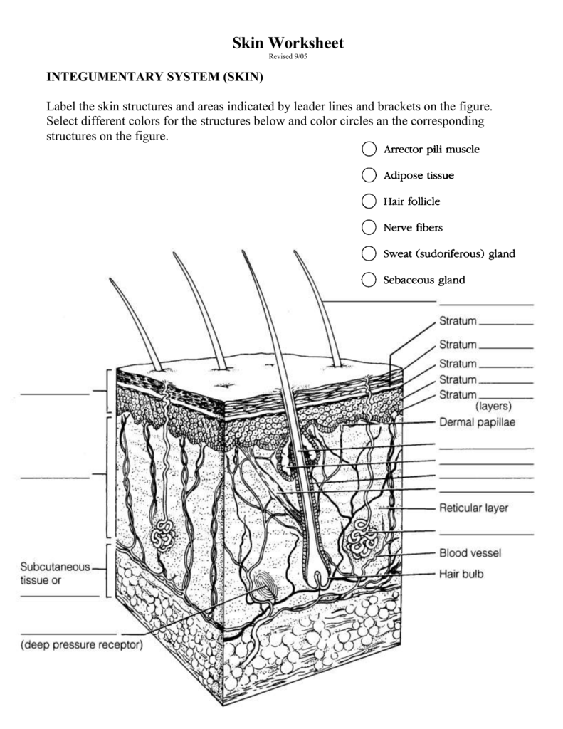
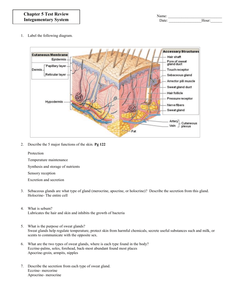
Layers of the Skin | Anatomy and Physiology I - Lumen Learning The skin is composed of two main layers: the epidermis, made of closely packed epithelial cells, and the dermis, made of dense, irregular connective tissue that houses blood vessels, hair follicles, sweat glands, and other structures. Beneath the dermis lies the hypodermis, which is composed mainly of loose connective and fatty tissues. Solved tive tissue 4. Label the skin structures and areas - Chegg Label the skin structures and areas indicated in the accompanying diagram of thin skin. Then, complete the statements that follow. Weisshaft Stratum opidamist Stratum Stratum Stratum Papilary layer Dermis Reticular layer ascectors allmusde Encrine Sweet blond Dermal Vascular plexus pensery neare fiber Blood vessel Subcutaneous tissue or Solved > 11.The terms and phrases in the key relate:1608365 ... - ScholarOn 4.Label the skin structures and areas indicated in the accompanying diagram of skin.... 5.What substance is manufactured in the skin (but is not a secretion) to play a role elsewhere in the body? ___________ 6.Some injections hurt more than others.... The Integumentary System - gserianne.com Label the skin structures and areas indicated in the accompanying diagram of thin skin. Then, complete the statements that follow. Subcutaneous tissue or.
Houston Community College Label the skin structures and areas indicated in the accompanying diagram of thin skin. Then, complete the statements that follow. Subcutaneous tissue or (deep pressure receptor) the epidermis. Stratum Stratum Stratum Stratum (layers) Papillary layer Reticular layer Blood vessel Adipose cells Solved > 3. Using the key choices, choose all responses:1607436 ... 1.layer of translucent cells in thick skin containing dead keratinocytes 2.two layers containing dead cells 3.dermal layer responsible for fingerprints 4.vascular region of the skin 5.major skin area as a whole that produces derivatives (nails and hair) 6.epidermal layer exhibiting the most rapid cell division SU_BIO1012_W3_A2_G1_Exercise7_HOLLOMAN_TUNISHA - Copy.pdf 45Basic Structure of the Skin 1. Complete the following statements by writing the appropriate word or phrase on the correspondingly numbered blank:The Integumentary System The two basic tissues of which the skin is composed are dense irregular connective tissue, which makes up the dermis, and 1, which forms the epi-dermis. Skin Structure (Labeling) Flashcards - Quizlet Start studying Skin Structure (Labeling). Learn vocabulary, terms, and more with flashcards, games, and other study tools.
Assignment 11 pg 104.pdf - 4. Label the skin structures and ...
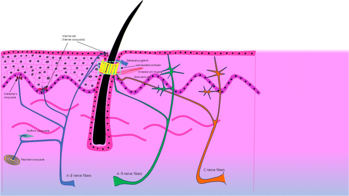
4. Label the skin structu - YUMPU Label the skin structures and areas indicated in the accompanying diagram of skin. epidermis dermis Subcutaneous tissue or hypodermis Pacinian corpuscle (deep pressure receptor) 5. What substance is manufactured in the skin (but is not a secretion) to play a role elsewhere in the body? The skin is the site of vitamin D synthesis for the body.
Regeneration of skin appendages and nerves: current status ...
PDF Skin Diagram Worksheet - uploads.strikinglycdn.com Nov 26, 2020 — Label the diagram in the spaces provided. ... What happens in the skin when blood vessels dilate, and how does this regulate temperature?. The two basic tissues of which the skin is composed are dense irregular ... Label the skin structures and areas indicated in the accompanying diagram of thin ....
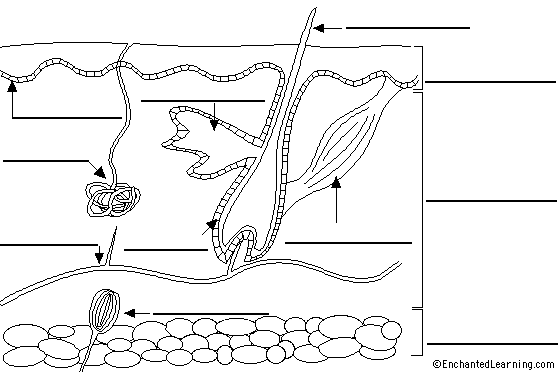
Label Skin Diagram Printout - EnchantedLearning.com
The Skin (Human Anatomy): Picture, Definition, Function, and Skin ... The skin is the largest organ of the body, with a total area of about 20 square feet. The skin protects us from microbes and the elements, helps regulate body temperature, and permits the...
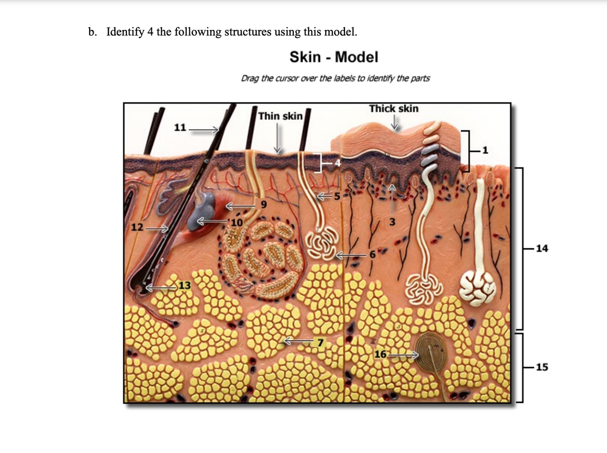
Answered: b. Identify 4 the following structures… | bartleby
PDF Integumentary System Review Sheet Exercise 7 Answers - Wise Bread An outer layer of cells designed to provide protection Keratinized stratified squamous epithelium. hypodermis (subcutaneous layer), this is a layer of *fat located under the dermis of the skin....

Skin Structure (Labeling) Flashcards | Quizlet
Answered: PRE-LAB Activity 6: Exploring the… | bartleby Science Biology Q&A Library PRE-LAB Activity 6: Exploring the Structure and Function of the Ear 1. Use the list of terms provided to label the accompanying illustration of the ear. Check off each term as you label it. O auricle O incus O stapes cochlea O malleus O tympanic membrane O external auditory canal O semicircular canals O vestibule a b. 17 2.
Untitled
PDF Integumentary Review Packet Key - Home - Buckeye Valley OBJECTIVE 1 structure(s) of the in OBJECTIVE 2 6. The layer where the skin is thick, such as the palms of the hands and the soles of the feet, is 10 -12 OBJECTIVE 3 OBJECTIVE 4 OBJECTIVE 5 OBJECTIVE 6 develops from a gro OBJECTIVE 7 c. production of gray pigments. d. reduction of melanocyte activity. 4.

File:Skin anatomy.jpg - Wikimedia Commons
PDF Teacher Instructions - Scholastic access to an outdoor area before 10 a.m. and again between 12 p.m. ... 5th-Grade Extension Have students write an accompanying report about what the skin does, how the sun can affect the skin, and ... You will create a 3-D model of the structure of the skin. Use the checklist to label the diagram below and make sure your model has all the ...
Explainer: What is skin? | Science News Explores
Integumentary System Review Sheet Exercise 7 squamous epithelium. hypodermis (subcutaneous layer), this is a layer of *fat located under the dermis of the skin. helps to insulate the body and protects underlying muscles and other structures. LAB Exercise 7 Integumentary System Questions and Study ... Start studying Review Sheet Exercise 7.

Living in Your Skin: Microbes, Molecules, and Mechanisms ...
PDF Review Sheet The Skin Integumentary System may 1st, 2018 - physio integumentary system review worksheet key objective describe the structure and functions of the skin 1 list the 5 functions of the integumentary system''CH4 SKIN LAB REVIEW SHEET ANSWERS PDF LEARNINGNETWORKS APRIL 30TH, 2018 - CREATED DATE 10 21 2013 12 25 55 PM'

Human skin - Wikipedia
Label the Skin | Biology Diagram | Quizlet Epidermis. layer of the skin that is continually being shed to make room for new skin. Dermis. layer of the skin that contains nerves, vessels, hair follicles, glands, and muscles. Hypodermis. layer of the skin that is used for fat storage. Sweat Pore. opening in the skin that sweat comes out of. Hair Follicle.
Plants | Free Full-Text | Ethnobotanical Uses, Phytochemical ...
Label Skin Diagram Printout - EnchantedLearning.com epidermis - the outer layer of the skin. hair follicle - a tube-shaped sheath that surrounds the part of the hair that is under the skin. It is located in the epidermis and the dermis. The hair is nourished by the follicle at its base (this is also where the hair grows). hair shaft - The part of the hair that is above the skin.

Skin Structure (Labeling) Flashcards | Quizlet
PDF 1.Base your answer to the following question on Which 3.The diagram ... Base your answers to questions 34 and 35 on the information and diagram below and on your knowledge of biology. A small water plant (elodea) was placed in bright sunlight for five hours as indicated below. Bubbles of oxygen gas were observed being released from the plant. A)producing sugar B)making protein

Quentin Bone. 17 August 1931—6 July 2021 | Biographical ...
Muscle System Label Answers - thesource2.metro.net Label the skin structures and areas indicated in the accompanying diagram of thin skin. Then, complete the statements that OCR GCSE (9-1) Physical Education J587/01 Physical factors … Label the muscle group A and the bone B. ... Compare the changes in the respiratory system of Dancer A to Dancer B ...
The Integumentary System
Lab_Ex._6_Review_Sheet_Answers0001.pdf Label the skin structures and areas indicated in the accompanying diagram of skin. -Hair shaft. REPIDERMIS .. Stratum Luci dum. Stratum CORNEum.
Genetics of Skin Cancer (PDQ®)–Health Professional Version - NCI
Integumentary System - Building a Medical Terminology Foundation The skin and accessory structures are the largest organ system in the human body. The integumentary system refers to the skin and its accessory structures. In the adult human body, the skin makes up about 16 percent of body weight and covers an area of 1.5 to 2 m 2. In fact, the skin and accessory structures are the largest organ system in the ...
Immune regulation by fungal strain diversity in inflammatory ...
Assignment 11 pg 104.pdf - 4. Label the skin structures and... Label the skin structures and areas indicated in the accompanying diagram of thin skin. Then, complete the statements that follow. Subcutaneous J tissue or _l T~P-r
The Integumentary System
Living Environment - New York Regents June 2009 Exam - Syvum Answer: 13. 13 Carbon dioxide makes up less than 1 percent of Earthâ s atmosphere, and oxygen makes up about 20 percent. These percentages are maintained most directly by (1) respiration and photosynthesis (2) the ozone shield (3) synthesis and digestion (4) energy recycling in ecosystems. Answer:
Protective effects of gallocatechin gallate against ...

excercise 7 p2.jpeg - 104 Review Sheet 7 4. Label the skin ...

Living in Your Skin: Microbes, Molecules, and Mechanisms ...

Polymeric Tissue Adhesives | Chemical Reviews
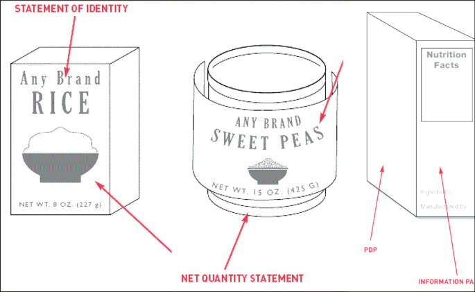
Food Labels | SpringerLink
Skin 1: the structure and functions of the skin | Nursing Times

Screen Shot 2020-08-26 at 12.21.00 AM.png - 4. Label the skin ...
BY ORDER OF THE SECRETARY OF THE AIR FORCE AIR FORCE PAMPHLET ...

Terahertz radiation and the skin: a review

SUSTAINABILITY COMMITMENT FOR INDONESIA
Label the Skin Anatomy Diagram

PDF) Neurocosmetics in Skincare—The Fascinating World of Skin ...
Skin Worksheet

Bioactive functional scaffolds for stem cells delivery in ...

Distinct skin morphological and transcriptomic profiles ...

Atopic dermatitis: Role of the skin barrier, environment ...

The emerging role of nanotechnology in skincare - ScienceDirect
Label a diagram of the skin - Mrs. Sanborn's Science Class

In situ visualization of glucocerebrosidase in human skin ...
biosphere | Definition, Resources, Cycles, Examples, & Facts ...

Skin Cancer Treatment (PDQ®) - PDQ Cancer Information ...

Superiority of microemulsion-based hydrogel for non-steroidal ...
A New Concept of Static Rubber Gasket for Sealing Rough Surface
Post a Comment for "40 label the skin structure and areas indicated in the accompanying diagram of skin"