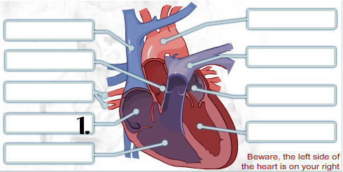
38 can you label this diagram of a human heart?
Human Heart Diagram - Human Body Pictures - Science for Kids Find free pictures, photos, diagrams, images and information related to the human body right here at Science Kids. Photo name: Human Heart Diagram Picture category: Human Body Image size: 70 KB Dimensions: 600 x 600 Photo description: This is an excellent human heart diagram which uses different colors to show different parts and also labels a number of important heart component such as the ... Label the heart - Science Learning Hub Label the heart Interactive Add to collection In this interactive, you can label parts of the human heart. Drag and drop the text labels onto the boxes next to the diagram. Selecting or hovering over a box will highlight each area in the diagram. Right ventricle Right atrium Left atrium Pulmonary artery Left ventricle Pulmonary vein Semilunar valve
Human Heart - Anatomy, Functions and Facts about Heart The human heart is located between the lungs in the thoracic cavity, slightly towards the left of the sternum (breastbone). It is derived from the embryonic mesodermal germ layer. The Function of Heart The function of the heart in any organism is to maintain a constant flow of blood throughout the body.

Can you label this diagram of a human heart?
How to Draw the Internal Structure of the Heart - wikiHow To draw the internal structure of a human heart, follow the steps below. Part 1 Finding a Diagram 1 To find a good diagram, go to Google Images, and type in "The Internal Structure of the Human Heart". Find an image that displays the entire heart, and click on it to enlarge it. 2 Find a piece of paper and something to draw with. labeling the heart diagram Heart Diagram - 15+ Free Printable Word, Excel, EPS, PSD Template . heart diagram human template medical sample templates printable diagrams word excel. Heart human diagram anatomy worksheets. Human anatomy worksheets. De heart anatomy diagram label mai in this interactive you can label Human Heart - Diagram and Anatomy of the Heart - Innerbody The heart is a muscular organ about the size of a closed fist that functions as the body's circulatory pump. It takes in deoxygenated blood through the veins and delivers it to the lungs for oxygenation before pumping it into the various arteries (which provide oxygen and nutrients to body tissues by transporting the blood throughout the body).
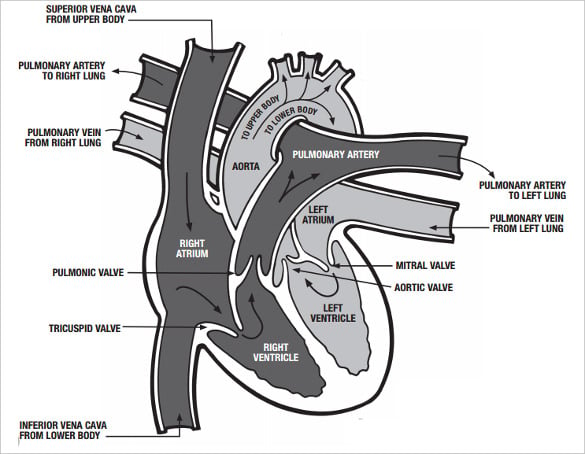
Can you label this diagram of a human heart?. Human Heart Diagram Labeled - Science Trends Let's examine the anatomy of the heart along with some diagrams that show how the heart operates. Anatomy Of The Heart The human heart usually weighs somewhere between 10 to 12 ounces in men and between 8 to 10 ounces in women, and in terms of size is roughly the size of the fist. Anatomy of a Human Heart - uofmhealth Located between the lungs in the middle of the chest, the heart pumps blood through the network of arteries and veins known as the cardiovascular system. It pushes blood to the body's organs, tissues and cells. Blood delivers oxygen and nutrients to every cell and removes the carbon dioxide and other waste products made by those cells. The Anatomy of the Heart - Quiz 1 - Free Anatomy Quiz The circulatory system - lower body image, with blank labels attached. The circulatory system - a PDF file of the upper and lower body for printing out to use off-line. Describe and explain the function of the circulatory system - The circulatory system consists of the heart, the blood vessels (veins, arteries, and capillaries), and the blood. Heart Labeling Quiz: How Much You Know About Heart Labeling? Here is a Heart labeling quiz for you. The human heart is a vital organ for every human. The more healthy your heart is, the longer the chances you have of surviving, so you better take care of it. Take the following quiz to know how much you know about your heart. Questions and Answers 1. What is #1? 2. What is #2? 3. What is #3? 4. What is #4?
Show me a diagram of the human heart? Here are a bunch! Diagram of a Labelled Heart with Direction of Blood Flow During Diastolic and Systolic Phases Human Heart: Direction of blood flow during Systolic and Diastolic Phases Realistic 3D Pictures of the Human Heart Oblique View of the Human Heart Heart myocardium and coronary vessels diagram Anatomy of a Normal Human Heart (anterior view) Labelling the heart — Science Learning Hub Blood transports oxygen and nutrients to the body. It is also involved in the removal of metabolic wastes. In this interactive, you can label parts of the human heart. Drag and drop the text labels onto the boxes next to the diagram. Selecting or hovering over a box will highlight each area in the diagram. Heart Diagram with Labels and Detailed Explanation - BYJUS The diagram of heart is beneficial for Class 10 and 12 and is frequently asked in the examinations. A detailed explanation of the heart along with a well-labelled diagram is given for reference. Well-Labelled Diagram of Heart The heart is made up of four chambers: The upper two chambers of the heart are called auricles. Solved: Label this diagram of the heart. | Chegg.com ISBN-13: 9780077350611 ISBN: 0077350611 Authors: Sylvia S Mader Rent | Buy. This is an alternate ISBN. View the primary ISBN for: Biology 10th Edition Textbook Solutions.
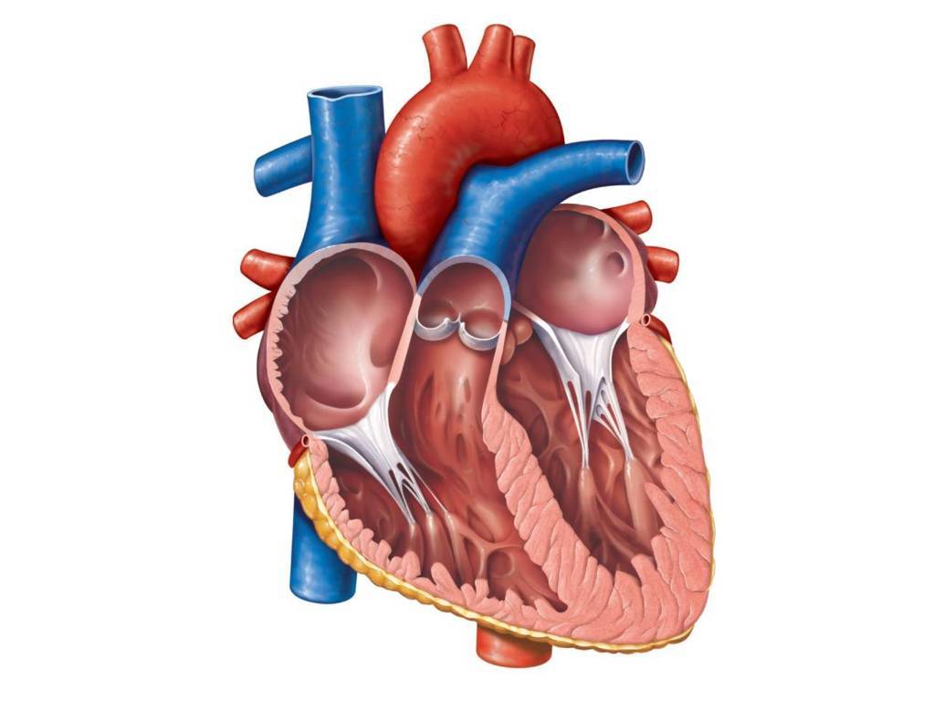
The Anatomy of the Heart, Its Structures, and Functions The heart is the organ that helps supply blood and oxygen to all parts of the body. It is divided by a partition (or septum) into two halves. The halves are, in turn, divided into four chambers. The heart is situated within the chest cavity and surrounded by a fluid-filled sac called the pericardium. This amazing muscle produces electrical ... Mastering Biology 8 Flashcards - Quizlet Can you label this diagram of a human heart? In what way can the human circulatory system be described as double circulation? The human cardiovascular system includes the pulmonary circuit and a systemic circuit. Oxygen-poor blood enters the heart through two veins: the superior vena cava and the inferior vena cava. ... How to Draw a Human Heart: 11 Steps (with Pictures) - wikiHow 3. Sketch a forked tube extending from the top of the rounded bump. To make the superior vena cava, draw a tube coming from the top of the right atrium. Make the tube fork about the same length as the bump you made for the right atrium chamber. Blood enters the right atrium through the superior vena cava. Heart Diagram - 15+ Free Printable Word, Excel, EPS, PSD Template ... This is a purely anatomical heart diagram for medical uses. Early 1911 Exposed Human Heart This is an antique presentation of a heart diagram on a thick card stock. This is a single sided diagram displayed in rustic color and industrial theme. It is perfect for anatomical use. Colour Heart Diagram
Animal Heart Diagram Labeled - ClipArt Best
Heart Anatomy: Labeled Diagram, Structures, Function, and Blood Flow Image: Cartoon diagram of the heart we will use to review the main cardiac structures and anatomy. Chambers of the Heart Let's begin with the chambers of the heart. There are 4 chambers, labeled 1-4 on the diagram below. To help simplify things, we can convert the heart into a square.
Heart Diagram – 15+ Free Printable Word, Excel, EPS, PSD Template ...
A Labeled Diagram of the Human Heart You Really Need to See A Labeled Diagram of the Human Heart You Really Need to See The heart, one of the most significant organs in the human body, is nothing but a muscular pump which pumps blood throughout the body. The human heart and its functions are truly fascinating. The heart, though small in size, performs highly significant functions that sustains human life.

Diagram of a Heart Labeled and Unlabeled 2018
Heart Dissection Questions - Heart Dissection Questions! Label the ... Label the diagram of the human heart below. Analysis Questions 1. How can you tell which side of the heart is the ventral surface (the surface closer to your chest)? By a groove that extends from the right side of the broad end of the heart diagonally to a point above & to your left of the apex. 1. Aorta2. Superior Vena Cava 3.

Label the heart — Science Learning Hub
Human Heart (Anatomy): Diagram, Function, Chambers, Location in ... - WebMD The heart is a muscular organ about the size of a fist, located just behind and slightly left of the breastbone. The heart pumps blood through the network of arteries and veins called the ...
Heart Quizzes, Trivia, Questions & Answers - ProProfs Quizzes
Solved Learning through Art: Blood Circulation through the | Chegg.com Biology questions and answers. Learning through Art: Blood Circulation through the Human Heart • Drag labels of Group 1 to trace the flow of oxygen-rich and oxygen poor blood. • Drag labels of Group 2 to identify the heart chambers. Reset Help Group 1 oxygen-poor blood from body Oxygen-rich blood Group 1 from lungs oxygen-rich blood to body ...

Human Heart Coloring Pages | Anatomy coloring book, Heart coloring ...
Heart Blood Flow | Simple Anatomy Diagram, Cardiac Circulation ... - EZmed Lastly we can revisit the original diagram shown at the beginning of this post and you should be able to understand and label the entire image. View fullsize Diagram: Blood flow through the heart, cardiac circulation pathway, and the main cardiac structures and anatomy.
How would you label the structures (both external and internal) of a ...
Diagram of Human Heart and Blood Circulation in It Exterior of the Human Heart A heart diagram labeled will provide plenty of information about the structure of your heart, including the wall of your heart. The wall of the heart has three different layers, such as the Myocardium, the Epicardium, and the Endocardium. Here's more about these three layers. Epicardium

A Diagram of the Heart and Its Functioning Explained in Detail
A Diagram of the Heart and Its Functioning Explained in Detail The heart blood flow diagram (flowchart) given below will help you to understand the pathway of blood through the heart.Initial five points denotes impure or deoxygenated blood and the last five points denotes pure or oxygenated blood. 1.Different Parts of the Body ↓ 2.Major Veins ↓ 3.Right Atrium ↓ 4.Right Ventricle ↓ 5.Pulmonary Artery ↓ 6.Lungs
Post a Comment for "38 can you label this diagram of a human heart?"