41 microscope diagram labeled
Cat Skeleton Anatomy with Labeled Diagram 29.5.2021 · Cat skeleton anatomy labeled diagram. Now, I will show you all the bones from the cat skeleton with a diagram. If you find any mistakes in this cat anatomy labeled diagram, please let me know. I hope this cat skeletal system anatomy labeled diagram might help you understand and identify all the cat’s bones. Microscope Diagram Labeled, Unlabeled and Blank | Parts of a Microscope ... This PDF contains the following: 1. Parts of a Microscope Diagram - Color 2. Parts of a Microscope Diagram - Black and White 3. Blank Parts of a Microscope Diagram - Black and White 4. Blank, Unlabeled Parts of a Microscope Diagram - Black and White 5. Blank Parts of a Microscope Diagram - Color 6. Blank, Unlabeled…
Parts of a Simple Microscope - Labeled (with diagrams) A simple microscope is a very first type of microscope ever created. It consists of simple parts and performs simple functions. Although there are now many advanced microscope types, some applications may still demand the use of a simple microscope. In this article, we are going to discuss the parts and functions of a simple microscope.
Microscope diagram labeled
Parts of a Microscope Labeling Activity - Storyboard That Create a poster that labels the parts of a microscope and includes descriptions of what each part does. Click "Start Assignment". Use a landscape poster layout (large or small). Search for a diagram of a microscope. Using arrows and textables label each part of the microscope and describe its function. Copy This Storyboard* More options Compound Microscope Parts, Functions, and Labeled Diagram Compound Microscope Parts, Functions, and Labeled Diagram Parts of a Compound Microscope Each part of the compound microscope serves its own unique function, with each being important to the function of the scope as a whole. Microscope Diagram | Quizlet This test will help the student to identify the different parts and function of the microscope. Terms in this set (12) Arm Supports the tube and connects it to the base Eyepiece the lens at the top that you look through. They are usually 10X or 15X power. Base The bottom of the microscope, used for support Body tube
Microscope diagram labeled. Compound Microscope Labeled Diagram | Quizlet QUESTION. The total magnification of a specimen being viewed with a 10X ocular lens and a 40X objective lens is. 15 answers. QUESTION. a mosquito beats its wings up and down 600 times per second, which you hear as a very annoying 600 Hz sound. if the air outside is 20 C, how far would a sound wave travel between wing beats. 2 answers. Compound Microscope Parts - Labeled Diagram and their Functions - Rs ... Labeled diagram of a compound microscope Major structural parts of a compound microscope There are three major structural parts of a compound microscope. The head includes the upper part of the microscope, which houses the most critical optical components, and the eyepiece tube of the microscope. Parts of a microscope with functions and labeled diagram 19.4.2022 · Figure: Diagram of parts of a microscope. There are three structural parts of the microscope i.e. head, base, and arm. Head – This is also known as the body. It carries the optical parts in the upper part of the microscope. Base – It acts as microscopes support. It also carries microscopic illuminators. A Study of the Microscope and its Functions With a Labeled Diagram To better understand the structure and function of a microscope, we need to take a look at the labeled microscope diagrams of the compound and electron microscope. These diagrams clearly explain the functioning of the microscopes along with their respective parts. Man's curiosity has led to great inventions. The microscope is one of them.
rsscience.com › stereo-microscopeParts of Stereo Microscope (Dissecting microscope) – labeled ... Labeled part diagram of a stereo microscope Major structural parts of a stereo microscope. There are three major structural parts of a stereo microscope. The viewing Head includes the upper part of the microscope, which houses the most critical optical components, including the eyepiece, objective lens, and light source of the microscope. Microscope Parts and Functions Microscope Parts and Functions With Labeled Diagram and Functions How does a Compound Microscope Work?. Before exploring microscope parts and functions, you should probably understand that the compound light microscope is more complicated than just a microscope with more than one lens.. First, the purpose of a microscope is to magnify a small object or to … 22 Parts Of a Microscope With Their Function And Labeled Diagram 22 Parts Of a Microscope With Their Function And Labeled Diagram Microscope Description A microscope is a laboratory instrument used to examine objects that are too small to be seen by the naked eye. In other words, it enlarges images of small objects. researchtweet.com › microscope-parts-labeledMicroscope, Microscope Parts, Labeled Diagram, and Functions Jan 19, 2022 · The liquid sample comes next. To minimise evaporation and protect the microscope lens from sample exposure, a small square of clear glass or plastic (a coverslip) is placed on top of the liquid. 1. Collect a clean microscope slide and a coverslip (a thin piece of plastic covering). Fill the centre of the microscope slide with a drop or two of ...
Compound Microscope- Definition, Labeled Diagram, Principle, … 3.11.2021 · A standard Microscope has three to four Objective Lenses which range from 4X to 100X. Stage Clips are metal clips that held the slide in place. Arm and Base. The Arm connects the Body Tube to the base of the Microscope. The Base supports the Microscope and its where Illuminator. Illuminator and Stage. The illuminator is the light source for a ... MICROSCOPE DIAGRAM - Google Search Microscope Diagram. (microscopic) of or relating to or used in microscopy; "microscopic analysis"; "microscopical examination". magnifier of the image of small objects; "the invention of the microscope led to the discovery of the cell". An optical instrument used for viewing very small objects, such as mineral samples or animal or plant cells ... microbenotes.com › parts-of-a-microscopeParts of a microscope with functions and labeled diagram Apr 19, 2022 · Figure: Diagram of parts of a microscope. There are three structural parts of the microscope i.e. head, base, and arm. Head – This is also known as the body. It carries the optical parts in the upper part of the microscope. Base – It acts as microscopes support. It also carries microscopic illuminators. Structure of Fungal Cell (With Diagram) | Fungi Electron microscope studies reveal that chitin occurs as elongated variously oriented microfibrillar units. These are laid down in layers and form the basis of the structural rigidity of fungal cell walls. The microfibril layers generally run parallel to the surface. Associated with the microfibrillar components is the nonfibrillar material.
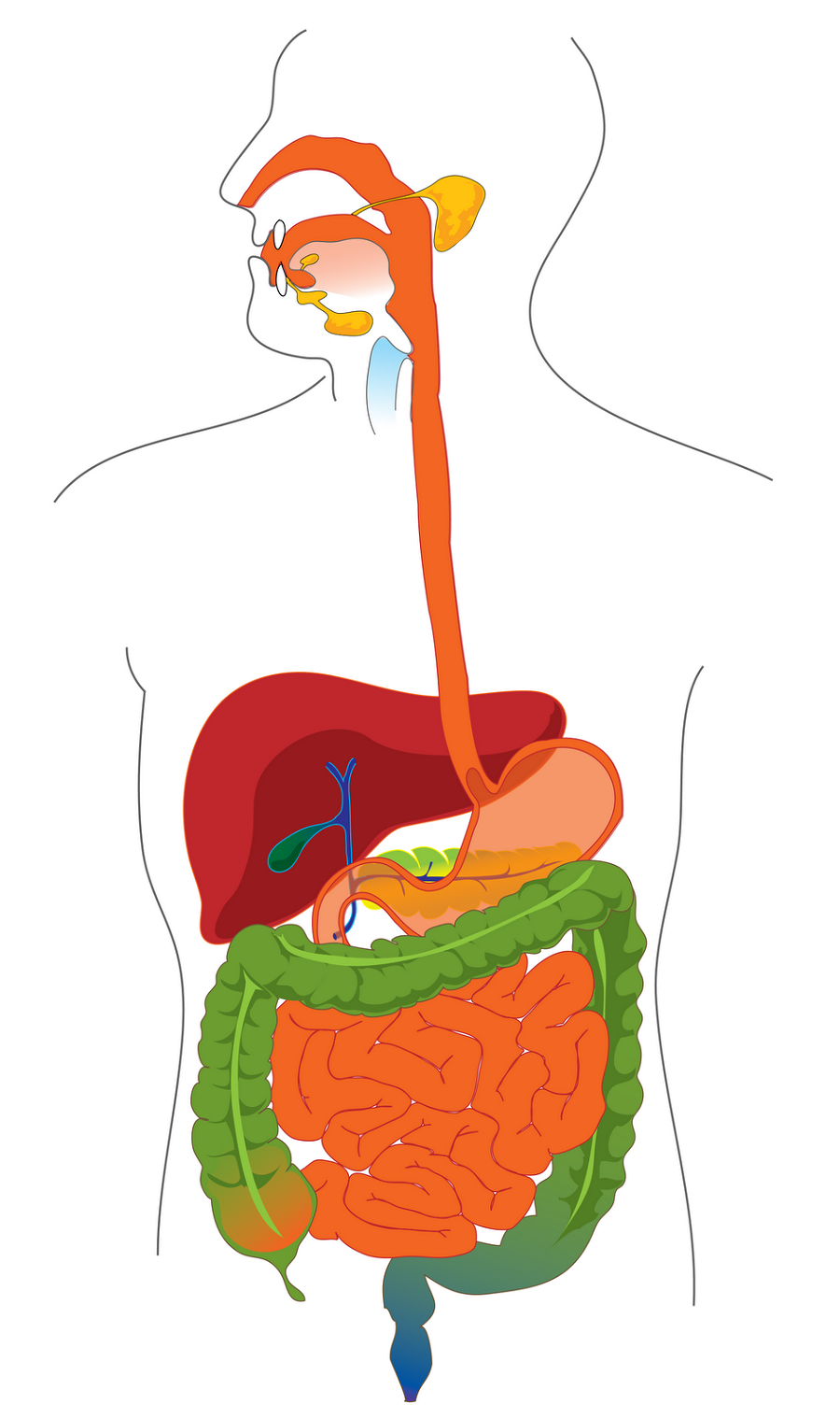
Diagram of the Digestive System
anatomylearner.com › cat-skeleton-anatomyCat Skeleton Anatomy with Labeled Diagram - AnatomyLearner May 29, 2021 · Cat skeleton anatomy labeled diagram. Now, I will show you all the bones from the cat skeleton with a diagram. If you find any mistakes in this cat anatomy labeled diagram, please let me know. I hope this cat skeletal system anatomy labeled diagram might help you understand and identify all the cat’s bones.
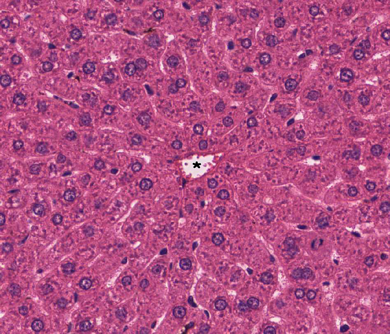
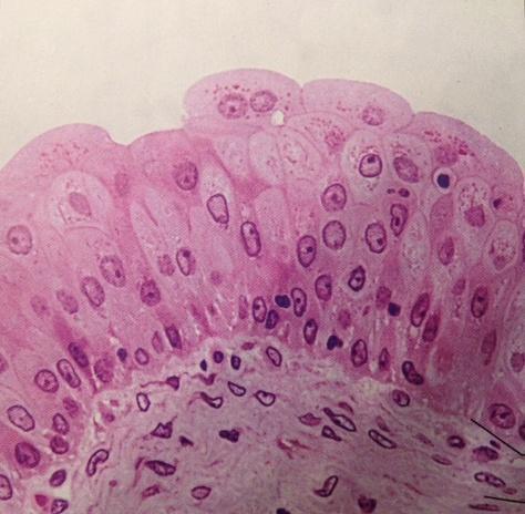
Liver, Gallbladder and Pancreas | histology
Parts of the Microscope with Labeling (also Free Printouts) 5. Knobs (fine and coarse) By adjusting the knob, you can adjust the focus of the microscope. The majority of the microscope models today have the knobs mounted on the same part of the device. Image 5: The circled parts of the microscope are the fine and coarse adjustment knobs. Picture Source: bp.blogspot.com.
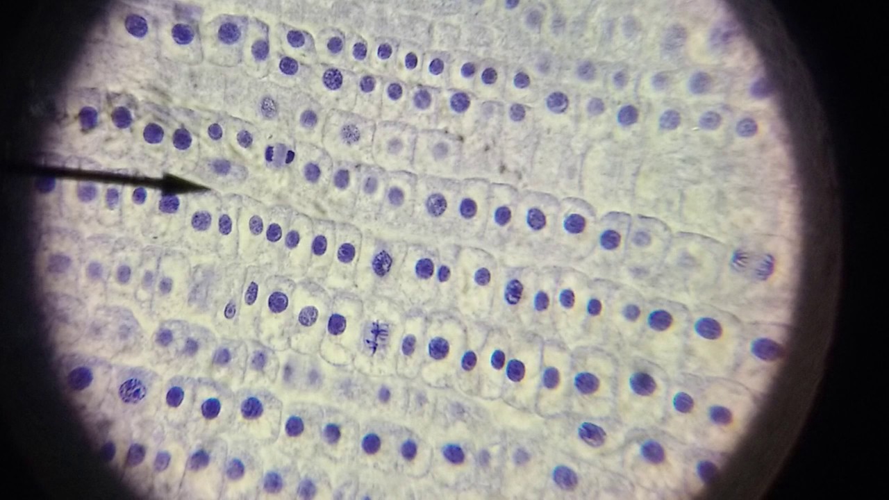
Mitosis in Onion Root Tips - YouTube
Labeling the Parts of the Microscope Labeling the Parts of the Microscope This activity has been designed for use in homes and schools. Each microscope layout (both blank and the version with answers) are available as PDF downloads. You can view a more in-depth review of each part of the microscope here. Download the Label the Parts of the Microscope PDF printable version here.

Onion peel cell diagram | Cell diagram, Diagram, Cell
Microscope Parts, Function, & Labeled Diagram - slidingmotion Microscope parts labeled diagram gives us all the information about its parts and their position in the microscope. Microscope Parts Labeled Diagram The principle of the Microscope gives you an exact reason to use it. It works on the 3 principles. Magnification Resolving Power Numerical Aperture. Parts of Microscope Head Base Arm Eyepiece Lens
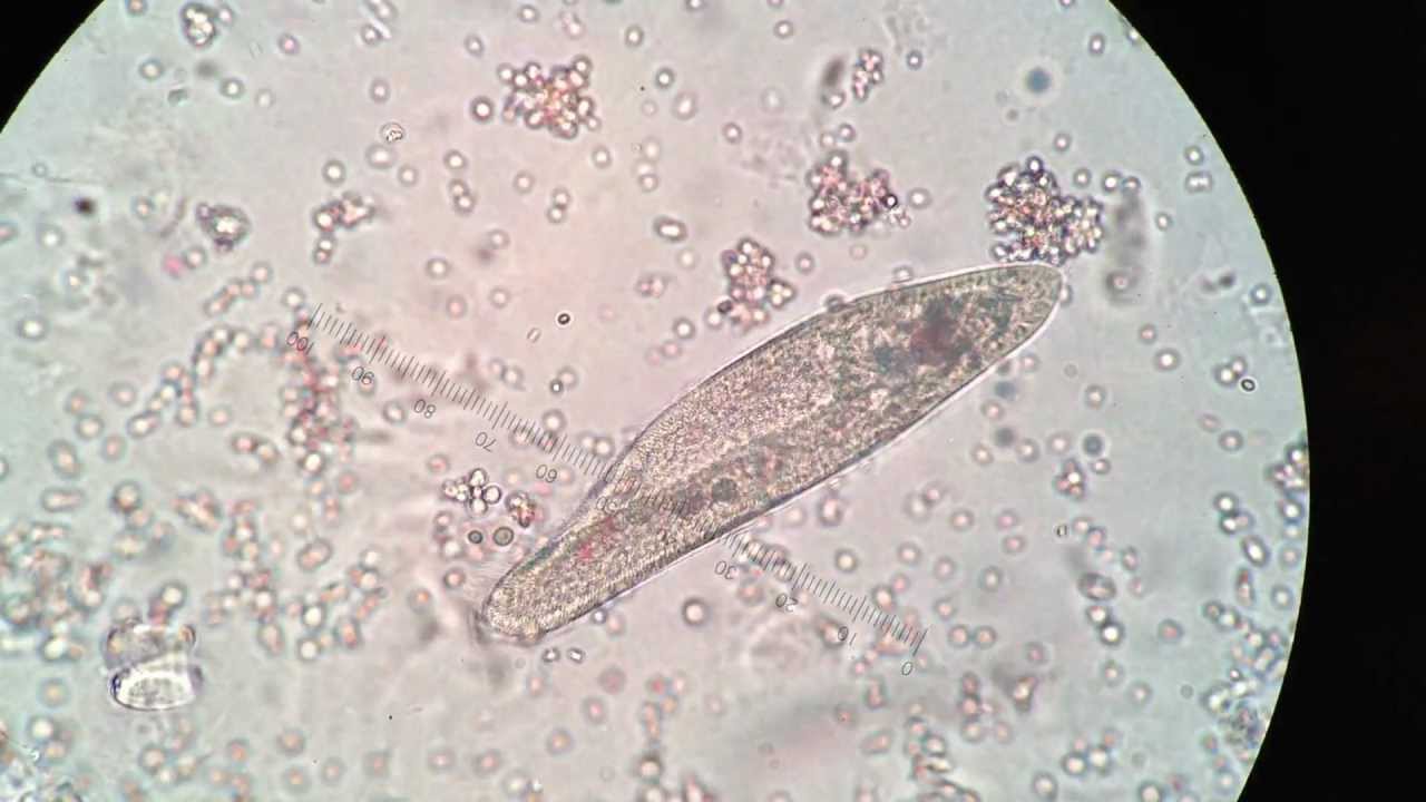
Paramecium under 400X magnification - YouTube
anatomylearner.com › ovary-histologyOvary Histology - Best Place to Learn Veterinary Anatomy Online May 02, 2021 · Ovary histology slide labeled diagram and drawing pictures. I am so happy to share ovary histology real slide picture, labeled diagram and drawing pictures with you. Hope these resources will help you lots to learn ovary histology. If you need more ovary real slide pictures then you may follow anatomy learner at here. Fallopian tube histology
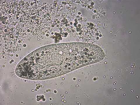
Paramecium under the microscope - YouTube
Parts of Stereo Microscope (Dissecting microscope) – labeled diagram ... If you would like to learn optical components of a compound microscope, please visit Compound Microscope Parts – Labeled Diagram and their Functions, and this article. How to use a stereo (dissecting) microscope. Follow these steps to put your stereo microscopes in work: 1.
Human Anatomy Lab Exercises Tissues Recognition and Function Flashcards ...
Microscope, Microscope Parts, Labeled Diagram, and Functions 19.1.2022 · Revolving Nosepiece or Turret: Turret is the part of the microscope that holds two or multiple objective lenses and helps to rotate objective lenses and also helps to easily change power. Objective Lenses: Three are 3 or 4 objective lenses on a microscope. The objective lenses almost always consist of 4x, 10x, 40x and 100x powers. The most common eyepiece …

Glomerulus histology of porcine kidney.... - Veterinary Medicine ...
Labelled Diagram of Compound Microscope - Biology Discussion The below mentioned article provides a labelled diagram of compound microscope. Part # 1. The Stand: The stand is made up of a heavy foot which carries a curved inclinable limb or arm bearing the body tube. The foot is generally horse shoe-shaped structure (Fig. 2) which rests on table top or any other surface on which the microscope in kept.

Histolab4ab.html
microbenotes.com › compound-microscope-principleCompound Microscope- Definition, Labeled Diagram, Principle ... Nov 03, 2021 · A standard Microscope has three to four Objective Lenses which range from 4X to 100X. Stage Clips are metal clips that held the slide in place. Arm and Base. The Arm connects the Body Tube to the base of the Microscope. The Base supports the Microscope and its where Illuminator. Illuminator and Stage. The illuminator is the light source for a ...
Post a Comment for "41 microscope diagram labeled"